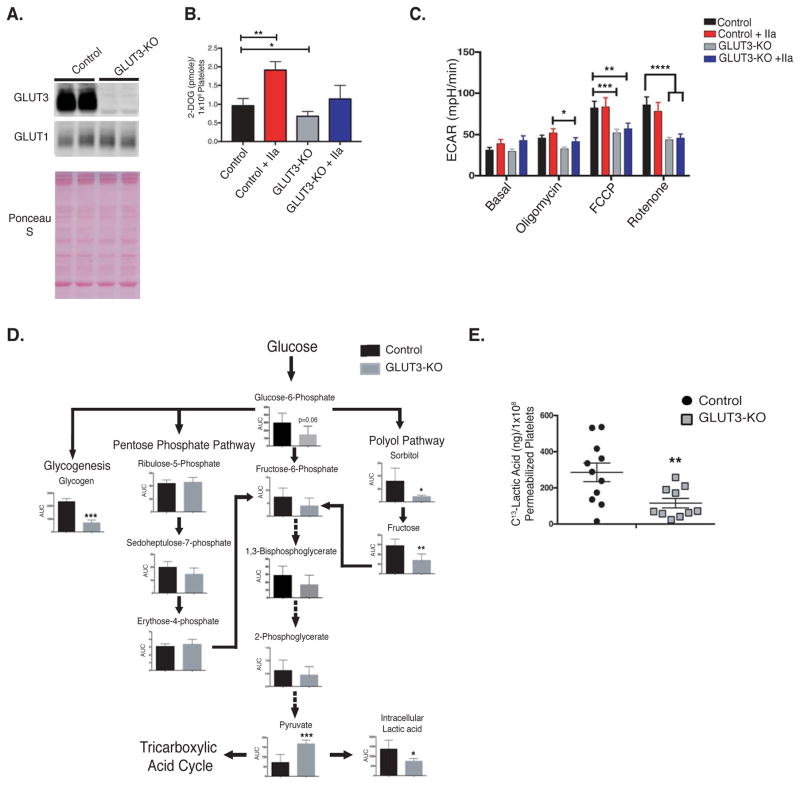
Figure 1. GLUT3 deletion reduces platelet glucose utilization.
(A) Western blot analysis of GLUT3 and GLUT1 protein in lysates from control and GLUT3 knockout platelets, n=6. (B) H3-2-deoxy-D-glucose (2-DOG) uptake in platelets was monitored in the presence or absence of 1U/mL thrombin (IIa), n=7. (C) Platelet glycolysis rate, as measured by the extracellular acidification rate (ECAR) was determined using a Seahorse XF24 analyzer. Platelet glycolysis rates with or without 1U/mL thrombin (IIa) were monitored under basal conditions and following treatment with the mitochondrial inhibitors: Oligomycin - ATP synthase inhibitor, Carbonyl cyanide-4 (trifluoromethoxy)phenylhydrazone (FCCP) – mitochondrial uncoupler, and rotenone, mitochondrial complex I inhibitor, n=5. (D) Metabolomics analysis of glycolytic intermediates in non-stimulated freshly isolated platelets were determined and normalized to platelet number, n=5. Glycogen analysis was determined fluorometrically by the enzymatic conversion of glycogen to glucose, n=6. (E) 13C-Lactic acid production by saponin permeabilized platelets, following incubation with 13C-Glucose, n=10. Data are mean±SEM. *P<0.05, **P<0.01,***P<0.001; 2-way ANOVA followed by Tukey’s multiple comparison post hoc test (B and C)); Student’s t test (D and E).

