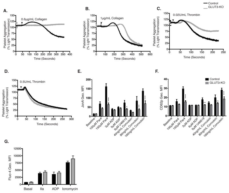
Figure 4. GLUT3 deletion leads to decreased platelet activation in vitro.
Representative tracings of washed platelets stimulated with (A) 0.6μg collagen, (B)1μg collagen (C) 0.02U/mL thrombin or (D) 0.5U/mL thrombin, at the indicated times (black arrowhead), n≥3. Diluted whole-blood stimulated with submaximal and maximal agonist concentrations. Platelets were monitored for GPIIbIIIa activation as marked by (E), JONA geometric mean fluorescence (Geo. MFI) and (F), CD62p Geo. MFI, n=6. (G) Platelets loaded with Fluo-4 were analyzed for basal and 1U/mL thrombin (IIa), 9μM ADP or 1μM ionomycin-stimulated cytoplasmic Ca2+ concentration, n=3. Data are mean±SEM. *P<0.05, **P<0.01, ***P<0.001, ****P<0.0001 vs. control same treatment; 2-way ANOVA followed by Tukey’s multiple comparison post hoc test (E, F, and G).

