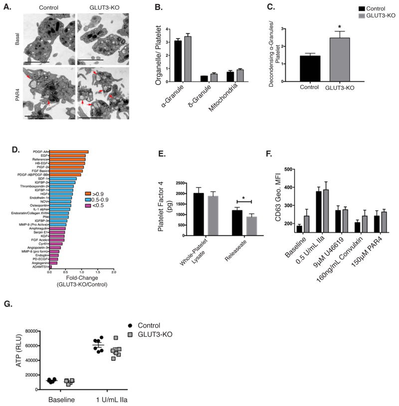
Figure 5. GLUT3 deletion leads to decreased α-granule degranulation in vitro.
(A) Transmission electron micrographs of washed platelets in suspension treated ± 250μM PAR4 peptide. α-granules in the process of degranulation (decondensing) depicted by red arrows, scale bar is 2μm. (B) Organelle density under non-stimulated conditions were quantified and normalized per platelet, n=4. (C) Quantification of α-granules in the process of degranulation (decondensing) in PAR4 stimulated platelets, n=4. (D) Par4 peptide stimulated platelet releasate was monitored using a targeted angiogenesis protein array and expressed as % change relative to control, n=1. (E) ELISA quantification of platelet factor 4 (PF4) in whole platelet lysates and releasate from platelets stimulated with thrombin (1U/mL), n=9. (F) The δ-granule maker CD63 binding (Geo. MFI) was monitored in washed platelets treated with the indicated agonist, n=3. (G) ATP release was monitored, n=6. Data are mean±SEM. *P<0.05 vs. control same treatment; 2-way ANOVA followed by Tukey’s multiple comparison post hoc test (B, E, F, and G). Student’s t test (C).

