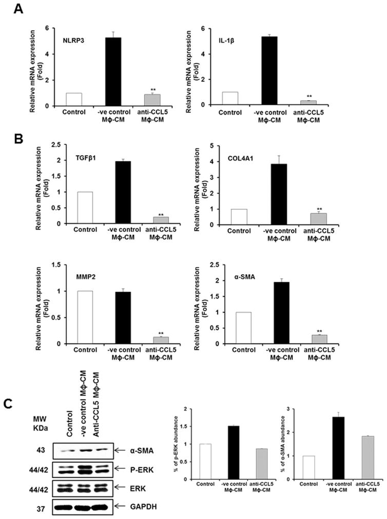
Figure 6. CCL5 from CM of HCV exposed macrophages induces inflammasome and profibrogenic markers.
Panel A. LX2 cells were incubated with HCV-MΦ-CM and CCL5 neutralizing antibody or negative control antibody (−ve cont Ab). Total RNA was isolated and qRT-PCR was performed to examine the expression of NLRP3 and IL-1β as described in figure 1. Panel B. LX2 cells were incubated with HCV-MΦ-CM and CCL5 neutralizing antibody or −ve cont Ab. Total RNA was isolated and qRT-PCR was performed for expression fibrogenic markers. Panel C. Cell lysates from above experiment were analyzed for expression of phosphor-ERK, total ERK and α-SMA by Western blot using specific antibodies. The blot was reprobed with antibody to GAPDH for loading control. Results from Densitometric scanning are presented in the right.

