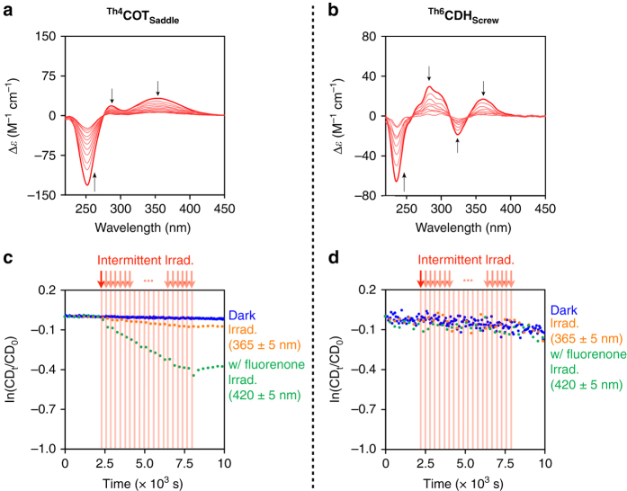
Fig. 3.
Ring inversion kinetics of Th4 COT Saddle and Th6 CDH Screw. a, b, Time-dependent CD spectral change profiles (240-s interval) of Th4 COT Saddle (a) and Th6 CDH Screw (b) in methylcyclohexane at 60 and 0 °C, respectively. Black arrows in a and b represent the directions of the CD spectral change with time. c, d, Decay profiles of the CD intensities at 260 nm (c, Th4 COT Saddle) and 280 nm (d, Th6 CDH Screw) in deaerated methylcyclohexane at 20 and −20 °C, respectively. Blue-colored dots represent decay profiles without photoirradiation. Orange-colored dots represent decay profiles with photoirradiation (λ = 365 ± 5 nm). Green-colored dots represent decay profiles with photoirradiation (λ = 420 ± 5 nm) in the presence of fluorenone (0.34 equiv. for Th4 COT Saddle and 0.23 equiv. for Th6 CDH Screw). Photoirradiation (250 s, red vertical lines) and CD spectroscopy (50 s, white area between red vertical lines) were conducted alternately for 6000 s

