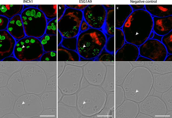
Figure 3.

INCh1 and ECG1A9 mAb labelling of starch granules in pea root cap. Images showing immunofluorescence labelling of starch granules by INCh1 (a) and ECG1A9 (b) in resin-imbedded sections through pea root caps. (c) No primary antibody control. Images shown in (a–c) are overlays of signals from three channels: Calcofluor White (staining cellulose, blue), propidium iodide (staining nuclei, red) and immunolabeling using an anti-mouse conjugated Alexa Fluor 488 as secondary antibody (green). The lower panels show differential interference contrast images obtained from the same sections. Starch granules are marked with arrowheads. Scale bars = 10 μm.
