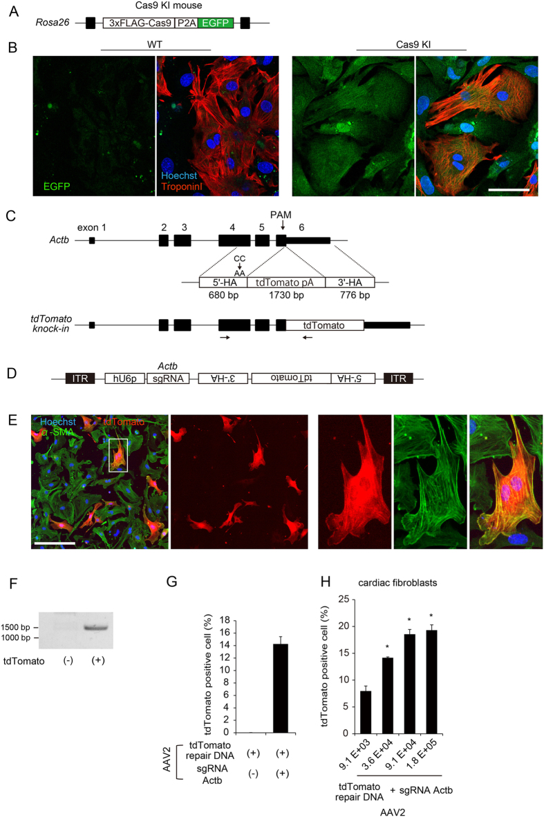
Figure 1.
Establishment of an evaluation method to detect HDR using a high-content image cytometry. (A) Cas9 knock-in mouse harbors a transgene encoding FLAG-Cas9-P2A-EGFP fusion protein at the Rosa26 locus. (B) Isolated cardiomyocytes and non-cardiomyocytes were cultured, fixed, and immunostained with anti–Troponin I antibody. EGFP signals were observed in both cell types. Scale bar: 50 μm. (C) The mouse Actb consists of 6 exons (upper). In the HDR repair template, the 1730-bp tdTomato fluorescent protein and 3′-terminal poly A sequence was cloned in-frame between the 680-bp 5′-terminal and 776-bp 3′-terminal homology arms (5′-HA and 3′-HA, respectively) corresponding to the genomic sequence of the 3′-terminus of Actb. PAM mutation (CC to AA) was introduced into 5′-HA, and stop codon sequence was removed. Expected genomic sequence of Actb-tdTomato fusion gene after successful HDR (lower). Arrows indicate locations of PCR primers (forward primer was designed outside homology arm). (D) Design of AAV vector. hU6 promoter (hU6p)-driven sgRNA and tdTomato HDR template including 5′-terminal homology arm (5′-HA) and 3′-terminal homology arm (3′-HA) were subcloned between inverse terminal repeat (ITR) sequences. (E) Cardiac fibroblasts isolated from Cas9 knock-in mice were seeded in 96-well plates and transduced with AAV serotype 2 (AAV2) encoding sgRNA and HDR template. Forty-eight hours after transduction, cells were fixed and stained with anti–α-SMA antibody. tdTomato-positive cell in white square is enlarged in right panels. Scale bar: 100 μm. (F) Cardiac fibroblasts were transduced with AAV as in (G). Forty-eight hours after transduction, genomic DNA were extracted from tdTomato-positive or –negative fibroblasts sorted by FACS. Genomic PCR was performed using a primer pair targeting inside and outside the homology arm (Fig. 1C). (H) Cardiac fibroblasts seeded in 96-well plates were transduced using AAV2 encoding HDR template with or without sgRNA targeting Actb. Forty-eight hours after transduction, cells were fixed and immunostained. The proportion of tdTomato-positive fibroblasts (among all cells) was calculated using the image cytometry (n = 3, means ± SD). (I) Cardiac fibroblasts seeded in 96-well plates were transduced using purified AAV in increasing titers (viral genomes per cell). Forth-eight hours after transduction, the proportion of tdTomato-positive fibroblasts were calculated as in (H). n = 3, *p < 0.01 vs low titer (9.1E + 03).

