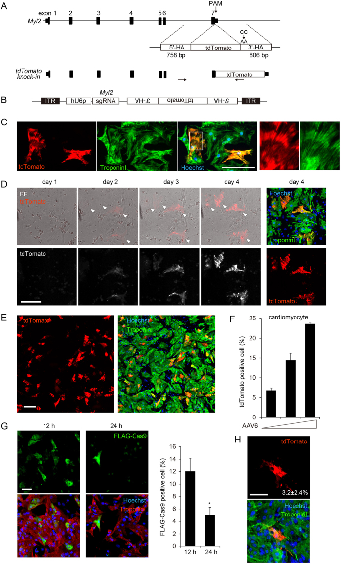
Figure 2.
Elucidation of the time course of HDR by sequential observation of individual cardiomyocytes. (A) The mouse Myl2 gene locus consists of 7 exons. HDR repair template consists of coding sequence of tdTomato fluorescent reporter protein between 758-bp 5′-terminal and 806-bp 3′-terminal homology arms (5′-HA and 3′-HA) corresponding to the genomic sequence of the 3′-terminus of Myl2. PAM mutation (CC to AA) was introduced into 3′-HA, and the stop codon sequence was removed. Arrows indicate the locations of PCR primers (outer primer was designed outside the homology arm). (B) Design of AAV vector. hU6 promoter (hU6p)-driven sgRNA and tdTomato HDR template were subcloned between inverse terminal repeat (ITR) sequences. (C) Cardiomyocytes isolated from Cas9 knock-in mice were seeded in 96-well plates and transduced with AAV serotype 6 (AAV6) encoding sgRNA and HDR template. Forty-eight hours after transduction, cells were fixed and stained with anti-troponin antibody. tdTomato-positive cardiomyocytes in white square are enlarged in right panels. Scale bar: 100 μm. (D) Cardiomyocytes were treated as in (C). After transduction of AAV, bright-field and fluorescence images were sequentially obtained using the image cytometry targeting the same fields determined by coordinate axes. On day 4, cells were fixed and immunostained. Arrowheads indicate the tdTomato-positive cardiomyocytes. Scale bar: 100 μm. (E) Immunostaining image of cardiomyocytes transduced with purified AAV6 encoding HDR components. Scale bar: 100 μm. (F) Cardiomyocytes were transduced with AAV6 at increasing viral titer (2.17 × 10⁴, 4.34 × 10⁴, and 1.09 × 10⁵ viral genomes/cell). At day 4, cells were fixed and immunostained. The proportion of tdTomato-positive cells among troponin I–positive cardiomyocytes was calculated using the image cytometry (n = 3, means ± SD). (G) Cardiomyocytes isolated from neonatal WT mice were transfected with mRNA encoding N-terminally FLAG-tagged Cas9. Twelve and 24 h after transfection, cells were fixed and immunostained. Scale bar: 50 μm. The proportion of FLAG-Cas9-positive cells among troponin I–positive cardiomyocytes was calculated using the image cytometry (n = 3, means ± SD, *: p < 0.05 vs 12 h). (H) WT cardiomyocytes were transduced with AAV6 (4.34 × 10⁴ viral genomes/cell). Twelve h after transduction, cardiomyocytes were transfected with mRNA encoding N-terminally FLAG-tagged Cas9. Four days after transfection, cells were fixed and immunostained. Scale bar: 50 μm. The proportion of tdTomato-positive cells among troponin I–positive cardiomyocytes was calculated using the image cytometry (n = 4, means ± SD).

