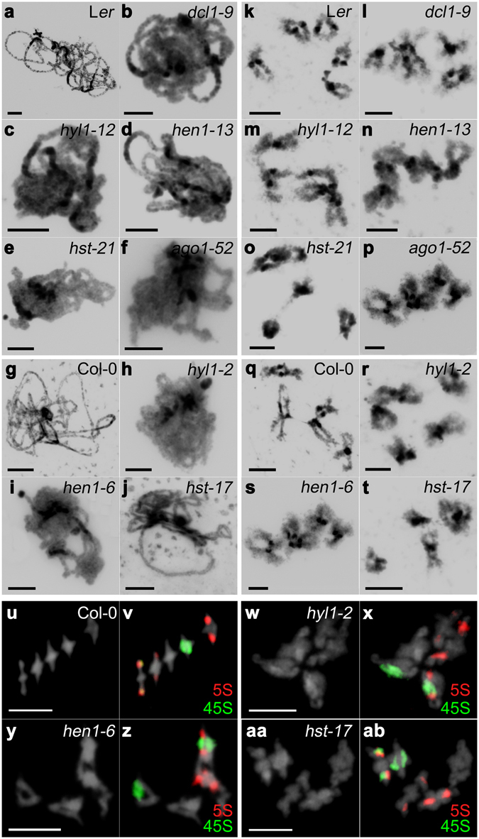
Figure 2.

Representative images of DAPI stained PMCs at pachytene and diakinesis, and FISH of metaphases I. (a–j) Pachytene. (k–t) Diakinesis. (u–ab) Diakinesis-Metaphase I. (a,k) Ler. (b,l) dcl1-9. (c,m) hyl1-12. (d,n) hen1-13. (e,o) hst-21. (f,p) ago1-52. (g,q,u,v) Col-0. (h,r,w,x) hyl1-2. (i,s,y,z) hen1-6. (j,t,aa,ab) hst-17. Mutant PMCs show a partial chromatin decondensation compared with wild type (see text for details). 5S rDNA is labeled in red and 45S rDNA in green. Bars = 5 µm.
