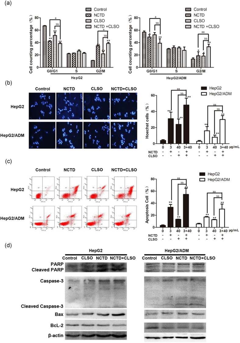
Figure 3.
Combination of NCTD and CLSO induces cell cycle arrest and apoptosis in HepG2 and HepG2/ADM cells. (a) Cell cycle distribution of HepG2 and HepG2/ADM cells was determined 24 h after treatment with NCTD and CLSO alone or in combination(n = 3). (b) Cells were stained with Hoechst33342 (5 μg/ml) and subjected to analysis of apoptosis population(n = 3). (c) PE-Annexin V staining of phosphatidylserine exposed on the cell surface was measured by flow cytometric analysis (n = 3). Data derived from three separate experiments are presented as the means ± S.D. (d) Total cell lysates were prepared for western blot analysis of the apoptosis regulatory proteins (n = 3). *P < 0.05; **P < 0.01, vs. control, aP < 0.05; aaP < 0.01 vs. NCTD alone, bP < 0.05; bbP < 0.01 vs. CLSO alone. One-way ANOVA, post hoc comparisons, Tukey’s test. Columns, means; error bars, SDs.

