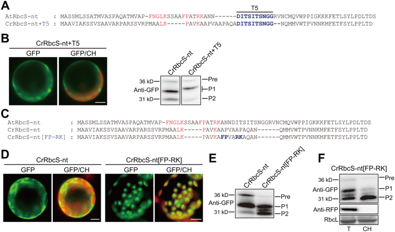
Figure 2.
The FP-RK motif of AtRbcS-nt rescues the defect of CrRbcS-nt in protein import into plant chloroplasts. (A and C) Sequences of AtRbcS TP, CrRbcS TP, and modified CrRbcS TP. All constructs were fused to GFP. (B and D) Localization of reporter proteins. Protoplasts from Arabidopsis plants were transformed with the indicated constructs, and GFP patterns were observed 12 h after transformation. Green, red, and yellow signals represent GFP, chlorophyll autofluorescence, and the overlap between green and red signals, respectively. Scale bar = 20 μm. In the western blot image of Fig. 2B, both lanes were cropped from the different parts of the same gel (Fig. S3). (E) Western analysis of reporter proteins. Total protein extracts from transformed protoplasts were analyzed by western blotting with anti-GFP antibody. Pre, precursor form; P1, processed form 1; P2, processed form 2. (F) Isolation of chloroplasts from transformed protoplasts. Chloroplasts were isolated from protoplasts transformed with CrRbcS-nt[FP-RK]:GFP. At 12 h after transformation, protoplasts were gently lysed and chloroplasts were isolated using a Percoll gradient. Total and chloroplast fractions were analyzed by western blotting using anti-GFP and anti-RFP antibodies. RFP was used as a control for cytosolic proteins. Rubisco complex large subunit (RbcL) stained with Coomassie brilliant blue was used as a loading control. T, total protein; CH, chloroplast fraction; Pre, precursor form; P1, processed form 1; P2, processed form 2.

