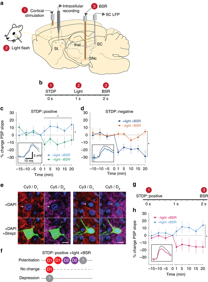
Fig. 2.

Reinforcement modulates corticostriatal STDP to enable bidirectional plasticity. a Schematic view of stimulating and recording sites. Numbers in red relate to components of the experimental protocol b. c Positive STDP pairings are modulated by the reward components (blue, n = 7) to induce lasting potentiation of corticostriatal synapses (time points after protocol vs. baseline, LME, est. effect size = 8.8 ± 3.3%, F 1,46 = 6.69, P = 0.013), which was significantly different from positive pairings without reinforcement (LME, est. effect size = 20.1 ± 8.8%, F 1,11 = 5.20, P = 0.043). Inset shows representative PSPs as defined in Fig. 1(f). d Negative STDP pairings are modulated by the reward components (dark blue, n = 5) to induce lasting depression of corticostriatal synapses, significantly different from negative pairings without reinforcement (LME, est. effect size = 24.5 ± 8.3%, F 1,10 = 10.40, P = 0.009). e Examples of D1 (Cy3) and D2 (Cy5) positive SPNs intracellularly filled with biocytin and visualized with DyLight 488-conjugated streptavidin. Note the presence of double labeling indicated by a perisomal ring within the cell membrane. Scale bar = 10 μm. f Summary dopamine receptor results for the positive STDP pairings with reinforcement group, indicating a mixed receptor expression in cells that potentiated. g Modified plasticity protocol without the light flash. h Positive STDP pairings, when modulated by BSR alone at 2 s (pink, n = 5), induce long-lasting depression, significantly different from positive pairings with light and BSR (LME, est. effect size = 24.4 ± 9.6%, F 1,10 = 6.40, P = 0.029). Data presented as mean ± s.e.m
