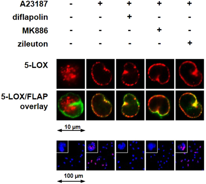
Figure 4.
Effect of diflapolin on 5-LOX subcellular redistribution and 5-LOX/FLAP interaction. Cells were pretreated with diflapolin (1 µM), MK886 (0.3 µM), zileuton (3 µM) or 0.1% DMSO for 15 min, and then stimulated with 2.5 µM Ca2+-ionophore A23187 for 10 min at 37 °C. Top panel: immunofluorescence microscopy was used to determine 5-LOX subcellular localization. Single images (top lane) show 5-LOX staining (red) and the overlay of 5-LOX (red) and FLAP (green) (middle lane). Results are representative for 100 individual cells of three independent experiments. Lower panel: in situ PLA was applied to determine 5-LOX/FLAP complex assembly in monocytes (lower panel) using antibodies against 5-LOX and FLAP. DAPI stains the nucleus (blue), and PLA signals by 5-LOX/FLAP complexes are stained in magenta. Results are representative for 100 individual cells of three independent experiments.

