
Figure 8.
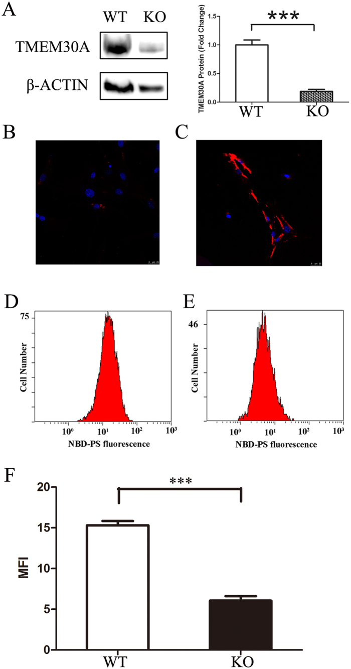
Loss of PS flippase activity in Tmem30a-mutant MEFs. (A) Western blot analysis revealed diminished Tmem30a expression in mutant MEFs. The expression of TMEM30A in mutant MEFs was reduced to 30% of that of control cells. Gel picture was cropped to save space. Full gel picture was listed as Figure S13. P<0.001. (B,C) Annexin V staining showed PS exposure on the mutant MEF cell surface, not on control cells. (D–F) NPD-PS labelling assay revealed loss of PS flippase activity in mutant MEFs. Internalization of NBD phospholipids by control (D) and mutant MEF cells (E). Loss of Tmem30a led to decreased NBD-PS uptake (E). The Y-axis showed numbers of NBD-PS-labelled MEF cells. The X-axis represented NBD-PS fluorescence intensity of intact living cells. Mutant cells exhibited decreased fluorescence intensity, compared to control cells (F). MFI, median fluorescence intensity. N = 3. ***P<0.001.