FIG 6.

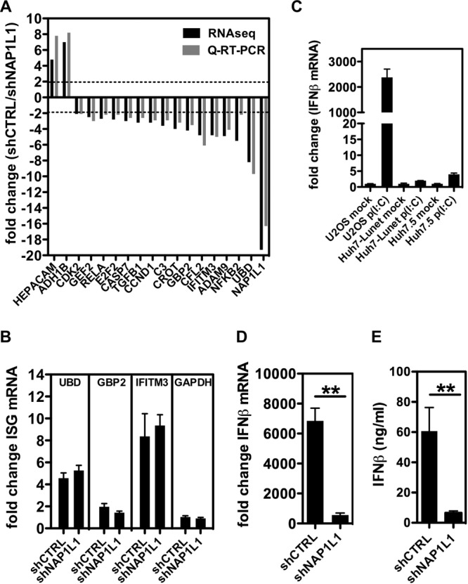
NAP1L1 is involved in the innate immune response. (A) Whole-genome transcriptome analysis in NAP1L1-depleted cells. Huh7-Lunet cells were treated with shNAP1L1/shCTRL, followed by RNA-seq analysis. Black bars show the levels of 15 downregulated genes (fold change, <−2) and 2 upregulated genes (fold change, >2), which were further validated by qRT-PCR (gray bars) normalized for β-actin. (B) Induction of ISGs is not NAP1L1 dependent. Huh7-Lunet cells were treated with 1000 U/ml of IFN-α for 8 h. UBD, GBP2, IFITM3, and GAPDH mRNAs were measured by qRT-PCR, normalized for β-actin in triplicate independent experiments. Shown are fold changes over basal, noninduced levels ± SD. (C) IFN induction following poly(I·C) transfection in different cell lines. U2OS, Huh7-Lunet, and Huh7.5 cells were transfected with 1 μg poly(I·C) for 8 h. IFN-β mRNA levels were measured by qRT-PCR, normalized for β-actin, and plotted against those for mock-treated cells (Lipofectamine). Average for 3 independent replicates are shown with standard deviations. (D) NAP1L1 depletion affects the induction of IFN-β mRNA by poly(I·C). U2OS cells treated with shNAP1L1/shCTRL for 3 days were transfected with 1 μg poly(I·C) for 8 h. IFN-β mRNA levels were measured as described above. (E) NAP1L1 depletion affects the induction of IFN-β by poly(I·C). U2OS cells were treated as for panel C. Secreted IFN-β protein was measured by a commercial enzyme-linked immunosorbent assay (ELISA) in triplicate and quantified against a standard curve, and results were plotted.
