FIG 7.
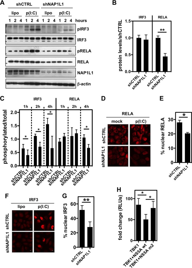
NAP1L1 controls RELA levels and IRF3 activation. (A) NAP1L1 depletion affects RELA levels and IRF3 phosphorylation. U2OS cells were transduced with LV for shNAP1L1 or shCTRL and subsequently transfected with 1 μg poly(I·C) using Lipofectamine (lipo). Protein levels as indicated were monitored by WB at 1, 2, and 4 h posttransfection of poly(I·C). (B) NAP1L1 depletion decreases RELA protein levels. Blots as in panel A were quantified to measure RELA and IRF3 protein levels using ImageJ. Shown is the shNAP1L1/shCTRL ratio in cells not transfected with poly(I·C). Averages for 3 independent replicates are shown with standard deviations. (C) NAP1L1 depletion affects IRF3 phosphorylation. Blots as in panel A were quantified to measure RELA and IRF3 phosphorylation levels using ImageJ. Shown is the ratio of phosphorylated protein to total protein in cells transfected with poly(I·C). Averages for 3 independent replicates are shown with standard deviations. (D) NAP1L1 depletion reduces RELA nuclear translocation. U2OS cells were transduced with LV for shNAP1L1 or shCTRL and subsequently transfected with 1 μg poly(I·C) for 8 h. Cells were then fixed and stained for RELA. (E) NAP1L1 depletion reduces RELA nuclear translocation. Around 500 cells from the experiment for panel D were counted for each condition to calculate the percentage of RELA nuclear translocation. Averages for 3 independent replicates are shown with standard deviations. (F) NAP1L1 depletion reduces IRF3 nuclear translocation. U2OS cells were transduced with LV for shNAP1L1 or shCTRL and subsequently transfected with 1 μg poly(I·C) for 8 h. Cells were then fixed and stained for IRF3. (G) NAP1L1 depletion reduces IRF3 nuclear translocation. Around 500 cells from the experiment for panel F were counted for each condition to calculate the percentage of IRF3 nuclear translocation. Averages for 3 independent replicates are shown with standard deviations. (H) HCV NS5A inhibits TBK1-mediated activation of IFN-β. HEK 293T cells were transfected with expression vectors for FLAG-tagged TBK1, NS5A, or the NS5A-m2 mutant together with a reporter plasmid carrying the firefly luciferase (Fluc) gene under the control of the IFN-β promoter (pIFNβ-Luc) and the control pCMV-Renilla. Relative light units (RLUs) of luciferase activity were measured in quintuplicate independent experiments, normalized for Renilla, and represented as fold change over mock treatment ± SD.

