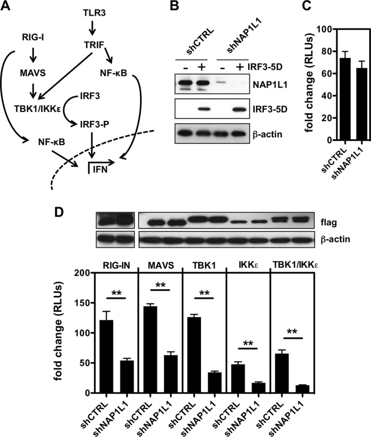
FIG 8.

NAP1L1 controls IRF3 phosphorylation at the TBK1/IKKε level. (A) Schematic representation of the RIG-I and TLR3 pathways. Both lead to activation of NF-κB and phosphorylation of IRF3 through MAVS/TBK1/IKKϵ or TRIF, respectively. NF-κB and pIRF3 translocate to the nucleus and activate IFN-β and other ISGs. (B) NAP1L1 does not affect constitutive IRF3-5D activity. Huh7-Lunet cells were transduced with LV for shNAP1L1 or shCTRL and subsequently transfected with an expression vector for IRF3-5D, the reporter IFN-β-Luc, and the Renilla control. Cell lysates were blotted as indicated. (C) NAP1L1 does not affect constitutive IRF3-5D activity. Luciferase activity of cells from the experiment for Fig. 7H was measured in triplicate independent experiments, normalized for Renilla. Average values are shown with standard deviations. (D) Depletion of NAP1L1 affects TBK1/IKKε-mediated activation of IFN-β. HEK 293T cells were transduced with LV for shNAP1L1 or shCTRL and subsequently transfected with expression vectors for FLAG-tagged RIG-I, IPS-1/MAVS, TBK1, and IKKε together with the reporter IFN-β-Luc and the Renilla control. Cell lysates were blotted with anti-FLAG antibody as indicated. Luciferase activity was measured in triplicate independent experiments, normalized for Renilla, and represented as fold change over mock treatment ± SD.
