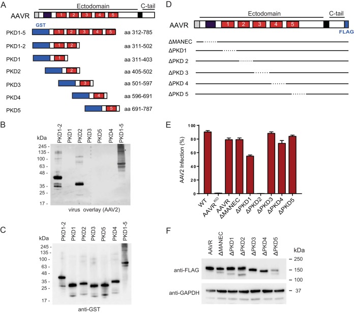
FIG 4.
AAV2 interacts with the second PKD domain in AAVR's ectodomain, and this interaction is critical for transduction. (A) Schematic depicting AAVR domains and various GST-tagged AAVR ectodomain constructs expressed in E. coli. (B and C) Gel electrophoresis (SDS–12% PAGE) was carried out on bacterial lysates of equal volumes of E. coli cells transformed with the respective constructs, and a virus overlay assay with rAAV2(Luc/mCherry) was performed (B), followed by reprobing with an anti-GST antibody (C). (D) Schematic depicting AAVR domains and deletion mutants, which remove a single domain per mutant. (E) scAAV2-CMV-RFP transduction of HeLa AAVRKO cells stably expressing AAVR deletion mutants depicted in panel D (MOI of 20,000 vg/cell). (F) Immunoblot of AAVR deletion mutants depicted in panel D, using anti-FLAG antibody and anti-GAPDH antibody. Cell pellets of 1 × 10⁶ cells were lysed with Laemmli SDS sample buffer containing 5% β-mercaptoethanol and were separated by SDS–4 to 15% PAGE, followed by immunoblotting. Data in panel E depict means with standard deviations from triplicate transductions, where transgene expression was measured after 48 h.

