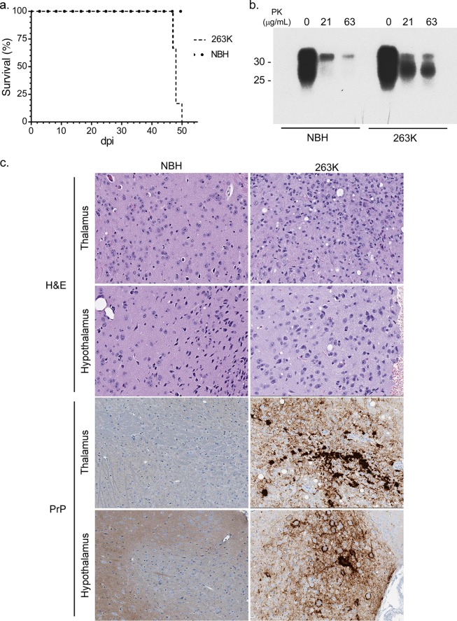
FIG 1.
Biochemical and histological examination of 263K hamster prion-infected Tg7 mice. (a) Survival curves for 263K hamster prion-infected (Tg7Sc) and normal brain homogenate-inoculated age-matched (Tg7NBH) Tg7 mice. The time to clinical disease was 48 ± 1.2 dpi for Tg7Sc mice (n = 6). Tg7NBH mice (n = 6) remained healthy for the duration of the experiment. (b) Brain tissue lysate from clinically ill (48 dpi) Tg7Sc mice and Tg7NBH was PK treated and probed with the anti-PrP mouse monoclonal 3F4 antibody. Tg7Sc mice were positive for protease-resistant PrPSc. Tg7NBH mouse samples were negative for PrPSc, with a nonspecific band at approximately 35 kDa. Molecular mass markers are shown on the left. (c) Sagittal brain sections from Tg7NBH and Tg7Sc mice were immunostained with the anti-PrP rabbit polyclonal antibody EP1802Y. Representative PrPSc staining and neuropathology in Tg7NBH (48 dpi) and Tg7Sc (48 dpi) mice are shown.

