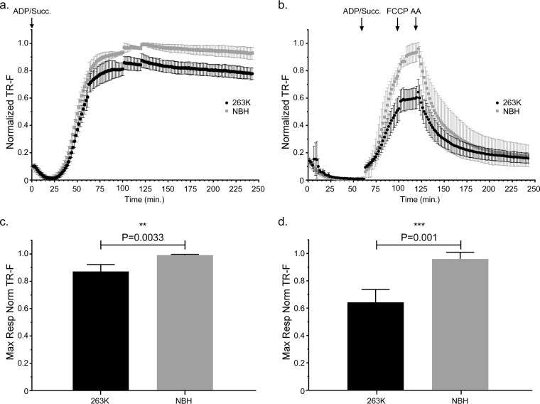
FIG 3.
Mitochondrial oxygen consumption is significantly decreased in clinically positive 263K prion-infected Tg7 mice. Isolated brain mitochondria from Tg7Sc mice euthanized at clinical stages of prion disease (48 dpi ± 1.2 dpi) or Tg7NBH-inoculated, age-matched control mice were assayed for their ability to take up oxygen in response to the complex II substrate succinate. (a) Saturating concentrations of ADP and succinate were added to each well at the beginning of the assay (arrow at 0 min, ADP/Succ.). Normalized fluorescence intensity of the oxygen reactive probe MitoXpress is shown. Data were normalized as described in the legend to Fig. 2. Error bars represent the normalized standard deviations for each time point. (b) Isolated brain mitochondria from Tg7NBH and Tg7Sc mice were allowed to equilibrate in the absence of substrate in order to obtain a basal level of oxygen uptake. Saturating concentrations of ADP and succinate (arrow at 60 min, ADP/Succ.) were added to wells containing mitochondria. The proton ionophore and oxidative phosphorylation uncoupler FCCP was then added to each well containing isolated mitochondria (arrow at 115 min, FCCP) to obtain maximal uncoupled respiratory rates. Lastly, the complex III inhibitor antimycin A was added (arrow at 135 min) to shut down electron transport. (c) Maximal respiration for panel a was determined by comparing the mean of the maximal values for each normalized value. (d) Maximal respiration for panel B was determined by comparing the mean of the maximal values for each normalized value. For panels c and d, an unpaired two-tailed Student t test was performed to determine significance. The normalized mean is derived from the average of 3 technical replicates for 4 experimental animals. Significance is denoted by the P value given above the bars.

