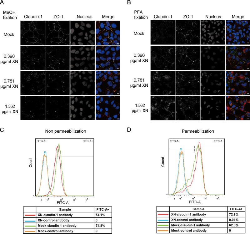
FIG 3.
Blocking COPII transport by XN decreased the cell surface level of claudin-1. (A) Huh7.5 cells treated with different concentrations of XN for 12 h were fixed with methanol and stained with antibodies against claudin-1 (red) and the tight junction marker ZO-1 (green). Nuclei were stained with DAPI (blue). Scale bar, 15 μm. (B) Huh7.5 cells treated with different doses of XN for 12 h were fixed with PFA and stained with antibodies against claudin-1 (red) and the tight junction marker ZO-1 (green). Nuclei were stained with DAPI (blue). Scale bar, 15 μm. (C) Cells surface claudin-1 was quantified by FACS assay in nonpermeabilized cells. Huh7.5 cells treated with 1.562 μg/ml XN or dimethyl sulfoxide (mock) for 12 h were harvested and stained with an anti-claudin-1 antibody (MAB4618; R&D Systems) without permeabilization. A total of 104 cells were analyzed, and isotype control antibody was used to delineate the gate. (D) Intercellular claudin-1 was quantified by FACS assay in permeabilized cells. Huh7.5 cells were treated with 1.562 μg/ml XN or dimethyl sulfoxide for 12 h. Then cells were permeabilized by saponin and stained with an anti-claudin-1 antibody (MAB4618; R&D Systems). A total of 104 cells were analyzed and isotype control antibody was used to delineate the gate.

