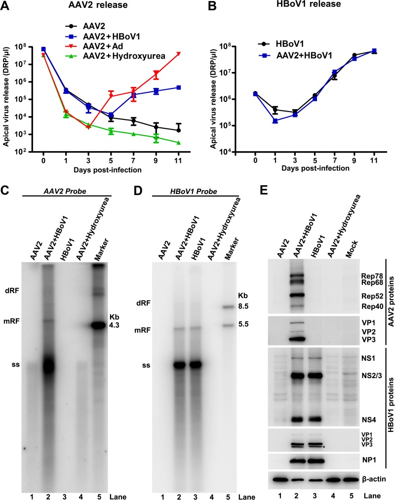
FIG 2.
HBoV1 rescues AAV2 replication in HAE-ALI cultures. (A) Virus release kinetics of AAV2. HAE-ALI cultures were infected with AAV2 in the presence of HBoV1 or Ad5 coinfection or treated with hydroxyurea at a final concentration of 2 mM. The released apical virions were collected every other day and measured by real-time PCR. Virus titers are shown as DRP per microliter. (B) HBoV1 virus release kinetics. HAE-ALI cultures were infected with HBoV1 alone or coinfected with AAV2. The amounts of HBoV1 virions released were measured by real-time PCR and are shown as DRP per microliter. (C and D) Southern blot analysis of AAV2 and HBoV1 DNA replication. Eleven days after infection or treatment with hydroxyurea, as indicated, 90% of the collected cells were lysed for Hirt DNA extraction. Hirt DNA samples were analyzed by Southern blotting with a 32P-labeled AAV2 probe (C) or a 32P-labeled HBoV1 probe (D). The dimer replicative form (dRF), monomer replicative form (mRF), and ssDNA (ss) are indicated. An AAV2 DNA at 4.3 kb (C) and HBoV1 DNA fragments at 5.5 kb and 8.5 kb (D) were used as size markers. (E) Western blot analysis of AAV2 and HBoV1 proteins. Eleven days after infection or treatment with hydroxyurea, as indicated, 10% of the collected cells were lysed and immunoblotted with anti-AAV2 Rep, anti-AAV2 VP, anti-HBoV1 NS1C, anti-HBoV1 VP3, anti-HBoV1 NP1, and anti-β-actin antibodies. Proteins detected are indicated next to the images. The asterisk indicates a possible cleaved VP3 protein.

