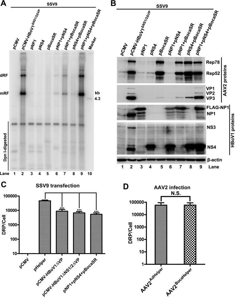
FIG 4.
AAV2 DNA replication, protein expression, and virus production in HEK293 cells cotransfected with different combinations of HBoV1 helper genes. HEK293 cells were transfected with an AAV2 infectious clone (SSV9) and various combinations of HBoV1 helper genes, as indicated. (A) Southern blot analysis. At 48 h posttransfection, 90% of the transfected cells were harvested for Hirt DNA extraction. Hirt DNA samples was examined for viral DNA replication by Southern blotting with a 32P-labeled AAV2 probe. dRF and mRF DNAs, DpnI-digested DNA, and the 4.3-kb AAV2 marker are indicated. (B) Western blot analysis. At 48 h posttransfection, 10% of the transfected cells were collected, lysed, and immunoblotted with anti-AAV2 Rep, anti-AAV2 VP, anti-HBoV1 NS1C, anti-HBoV1 NP1, and anti-β-actin antibodies. Proteins detected are indicated. (C) Production of AAV2. Transfected cells were collected, lysed, and quantified for DRP by real-time PCR. The virus production levels are shown as DRP per cell. Error bars show standard deviations, which were obtained from three independent experiments. Statistical analysis was performed by using the Student t test. ***, P < 0.0001. (D) Infectivity of the progeny viruses. HEK293 cells were infected with Ad pHelper-produced AAV2 (AAV2AdHelper) or with HBoV1 Helper-produced AAV2 (AAV2BocaHelper) at an MOI of 300 DRP/cell, followed by transfection with Ad pHelper. At 48 h posttransfection, the cells were collected for analysis of virus production, as determined by real-time PCR. Error bars show standard deviations, which were obtained from three independent experiments. Statistical analysis was performed by using the Student t test. N.S., no significance.

