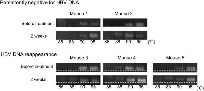
FIG 4.

HBV hypermutation following treatment with PEG-IFN and entecavir. HBV DNA was amplified by 3D-PCR, and hypermutated genomes were detected by HA-yellow agarose gel electrophoresis in five mice that were positive for serum HBV DNA after 2 weeks of treatment with PEG-IFN plus entecavir. Serum HBV DNA remained persistently negative in two mice (persistently negative for HBV DNA; mice 1 and 2) and reappeared after cessation of the treatment in three mice (HBV DNA reappearance; mice 3, 4, and 5). More heavily hypermutated HBV DNA was observed at 88°C. The dashed lines were added to help visualize the retardation of AT-rich DNA in the HA-yellow agarose gel.
