FIG 2.

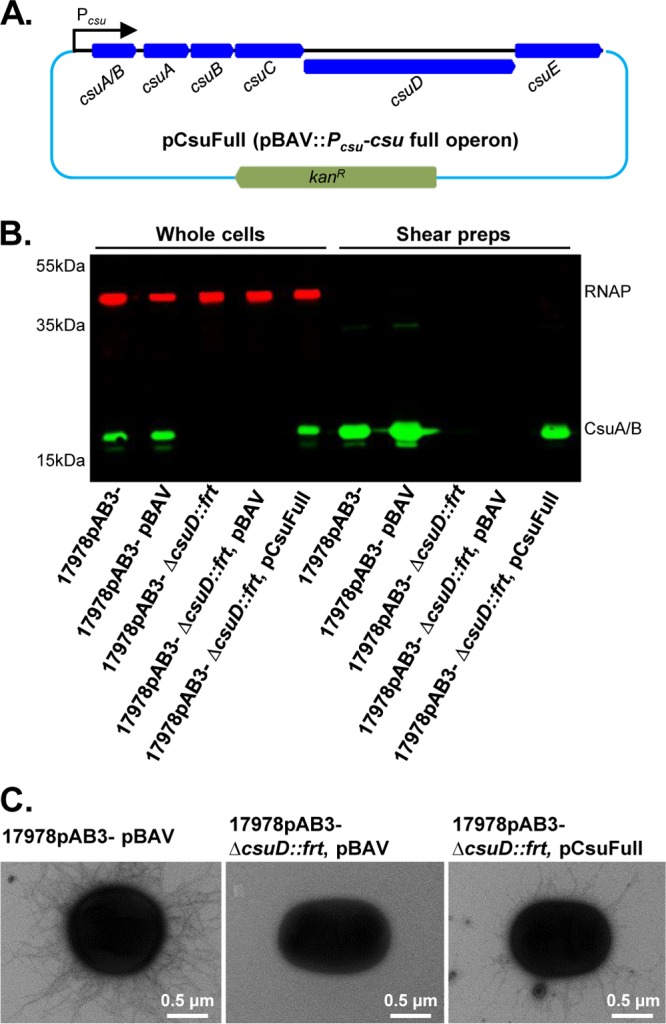
Complementation of Csu pili in 17978pAB3− ΔcsuD::frt. (A) Schematic diagram of the complementation plasmid pCsuFull (pBAV::csu full operon). The entire csu gene cluster, including its own promoter region, was cloned into the pBAV shuttle vector, which confers resistance to kanamycin. The pCsuFull plasmid was then electroporated in 17978pAB3− ΔcsuD::frt competent cells to complement the mutant in trans. (B) Confirmation of the expression of Csu pili in complemented cells by Western blotting. Total cell lysates and shear preparation samples were subjected to SDS-PAGE and immunoblotting with the anti-CsuA/B antibody (green bands); RNAP was used as a loading control (red bands). CsuA/B was detected in the 17978pAB3− wild-type strain and its vector control strain (17978pAB3− pBAV) but not in the 17978pAB3− ΔcsuD::frt strain and its vector control strain (17978pAB3− ΔcsuD::frt pBAV). The complemented strain (17978pAB3− ΔcsuD::frt pCsuFull) restored the CsuA/B expression. (C) Detection of Csu pili on the surface of the 17978pAB3− ΔcsuD::frt pCsuFull complemented strain. Transmission electron microscopic analysis confirmed the presence of Csu pili on wild-type cells (17978pAB3− pBAV) and complemented strain cells (17978pAB3− ΔcsuD::frt pCsuFull), whereas mutant cells (17978pAB3− ΔcsuD::frt pBAV) did not show Csu pili.
