FIG 9.

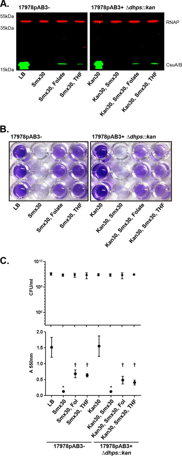
Recovery of Csu pilus and biofilm repression with both THF and folate supplements under Smx-mediated folate stress conditions. (A) CsuA/B expression with folate or THF supplements, as determined by Western blotting with the anti-CsuA/B antibody (green bands); RNAP was used as loading control (red bands). Folate stress was generated by Smx. Kan, kanamycin. (B) Representative crystal violet assay images of 17978pAB3− and 17978pAB3+ Δdhps::kan cells with folate or THF supplements. Both folate and THF supplements were able to relieve folate stress generated by Smx, resulting in partially restored biofilm formation. (C) CFU counts (top) and biofilm crystal violet assay quantification (bottom). Points indicate mean values, and error bars indicate standard deviations of triplicate samples. *, P ≤ 0.002; †, P ≤ 0.05, significant reduction of biofilm production, compared to 17978pAB3− cells with LB.
