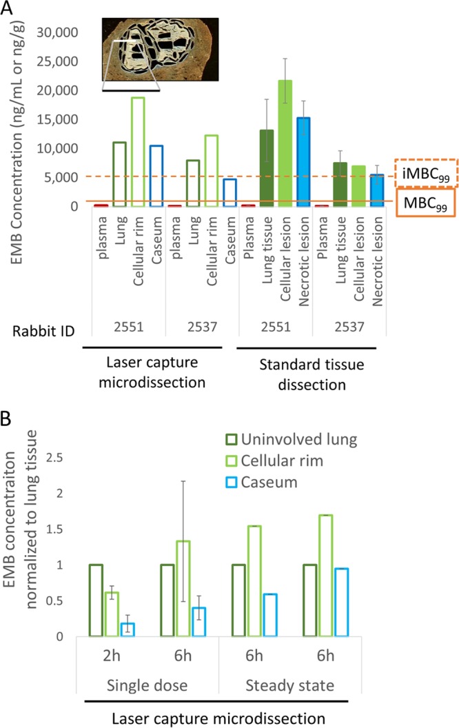
FIG 3.

Spatial quantitation of EMB in lung and lesion compartments. (A) The left side of the panel shows absolute EMB concentrations (open bars) measured by LC/MS-MS in lung and distinct regions of necrotic granulomas, laser captured and dissected from thin-tissue sections as shown at the top of the panel (see Fig. S3 in the supplemental material for the detailed procedure). The right half of the panel (filled bars) shows data acquired by LC/MS-MS in tissue homogenates collected by standard dissection of uninvolved lung, whole cellular lesions, and whole necrotic lesions. Both rabbits 2551 and 2537 received 100 mg/kg EMB daily for 7 days, and lesions were dissected 6 h after the last dose (steady state). The minimum concentrations required to kill 99% of extracellular replicating bacilli (MBC99) and 99% of intracellular bacilli in macrophages (iMBC99) are indicated (5, 30). (B) Comparison of EMB concentration ratios between lung and cellular or necrotic lesion compartments following a single dose and at steady state. Absolute EMB concentrations were measured by LC/MS-MS in uninvolved lung, cellular rim, and necrotic core of caseous granulomas, laser captured and dissected from thin tissue sections as shown in panel A.
