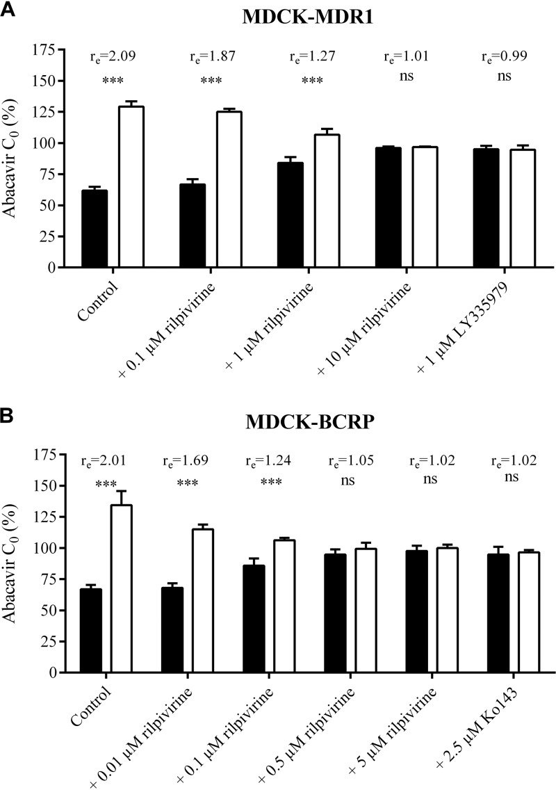
FIG 3.
Concentration equilibrium assay of [3H]abacavir (300 nM) in MDCK-MDR1 (A) and MDCK-BCRP (B) cells. The percentage of initial concentration (C0) of abacavir obtained in the basolateral (black columns) or apical compartment (white columns) after 6 h of incubation with or without rilpivirine is shown. LY335979 (1 μM) or Ko143 (2.5 μM) was used as a positive control. The ratio (re) between concentrations in the apical and basolateral compartments measured at the end of the experiment is shown. Data are shown as mean values ± SD from three experiments performed in duplicate. Statistical significance was analyzed by Student's t test. ***, P ≤ 0.001; ns, not significant.

