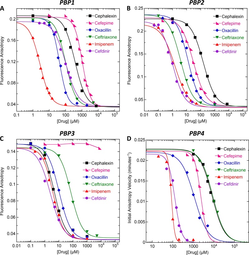
FIG 3.
(A to C) Fluorescence anisotropy of 1 μM Bocillin in the presence of the indicated β-lactam antibiotic and 2 μM of either PBP1 (A), PBP2 (B), or PBP3 (C). (D) Initial anisotropy velocity of 1 μM Bocillin in the presence of the indicated β-lactam and 50 μM PBP4. The solid lines reflect the nonlinear least-squares fits of the data with equation 1, with the exception of the curve for cefepime and PBP3, which could not be fit due to the weak binding of the drug. Experimental conditions were as described in the legends to Fig. 1 (for PBP1, PBP2, and PBP3) and Fig. 2 (for PBP4).

