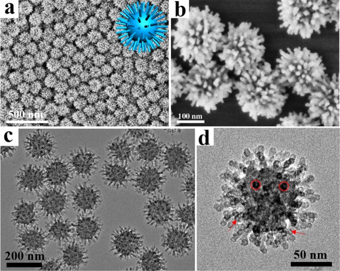
Figure 1.

Structural characterization of virus-like mesoporous silica nanoparticles. (a, b) SEM and (c, d) TEM images with different magnifications of the virus-like mesoporous silica nanoparticles. The red arrows mark the open tubular structures, and the red circles highlight the top view of the open silica nanotubes. The inset of (a) is the structural model for the virus-like mesoporous silica.
