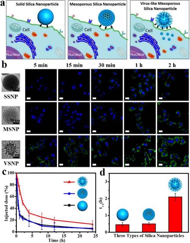
Figure 4.
Cellular internalization and in vivo blood circulation. (a) Schematic illustration for cellular uptake of three types of silica nanoparticles. (b) Confocal laser scanning microscopy (CLSM) observations of the HeLa cells after incubation with the solid silica nanoparticles (SSNPs), conventional mesoporous silica nanoparticles (MSNPs), and the virus-like mesoporous silica nanoparticles (VSNPs) for 5 min, 15 min, 30 min, 1 and 2 h. (c) Time-dependent blood level upon tail vein injection of three types of nanoparticles, calculated as percentage of injected dose remaining in the blood. (d) Blood circulation half-lives (t1/2) of three types of nanoparticles. Error bars were based on three mice per group at each time point and three repetitions, P < 0.05. All the scale bars are 10 μm.

