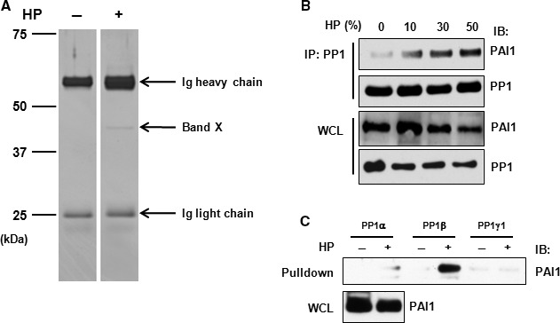
Figure 6.

Identification of PAI1 as a PP1‐interacting protein. (A) HCMECs were treated with or without HP for 30 min. according to Scheme B and whole‐cell lysates subjected to immunoprecipitation using an anti‐PP1 antibody followed by SDS‐PAGE separation and silver staining. The positions of molecular mass markers are marked on the left. Band X is the gel band of interest which was subjected to LC‐MS/MS analysis. (B) HCMECs were treated with HP for 30 min. and whole‐cell lysates were prepared and subjected to immunoprecipitation–immunoblotting. (C) Whole‐cell lysates prepared from HCMECs treated with or without HP were incubated with an equal molar amount of GST‐tagged PP1 isoforms followed by pull‐down with glutathione‐sepharose beads. The eluted proteins were resolved by SDS‐PAGE followed by immunoblotting. Ig, immunoglobulin; IP, immunoprecipitation; IB: immunoblotting; WCL: whole‐cell lysate.
