Figure 3.

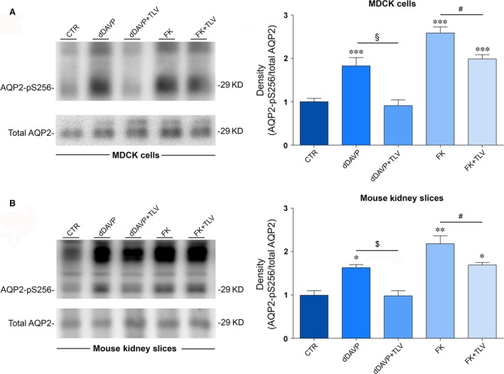
Effect of tolvaptan on AQP2 S256 phosphorylation in MDCK‐hAQP2 lysates and mouse kidney slices. (A) MDCK‐hAQP2 cells were treated as described under concise methods. Equal amount of proteins from cells (30 μg) were immunoblotted for total AQP2 and AQP2 phosphorylated at S256 (AQP2‐pS256). Signals were semiquantified by densitometry (right panel). Statistical analysis (means ± S.E.Ms; ***P < 0.0001 versus CTR; §P < 0.0001 dDAVP versus dDAVP+TLV; #P < 0.01 FK versus FK+TLV; n = 6) revealed that tolvaptan prevented the increase in AQP2‐pS256 compared with dDAVP and forskolin stimulation. (B) Mouse kidney slices were treated as described above. Equal amount of proteins from cells (15 μg) were immunoblotted for total AQP2 and AQP2 phosphorylated at S256 (AQP2‐pS256). Signals were semiquantified by densitometry (right panel). Statistical analysis (means ± S.E.Ms; *P < 0.01 versus CTR; **P < 0.001 versus CTR; $P < 0.01 dDAVP versus dDAVP+TLV; #P < 0.01 FK versus FK+TLV; n = 3) revealed that tolvaptan prevented the increase in AQP2‐pS256 compared with dDAVP and forskolin stimulation.
