Figure 1.

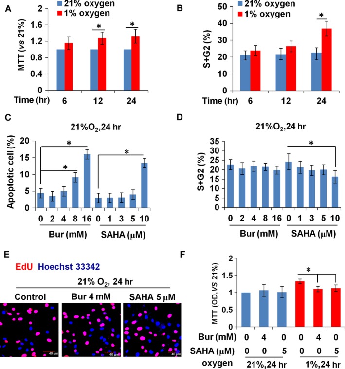
HDI at high concentrations inhibits the growth of VSMCs under normoxic conditions. (A) The proliferative activity of VSMCs, as detected by MTT assay, was increased significantly by hypoxia treatment for 12 and 24 hr. (B) Cell cycle analysis showed that the percentage of cells in the S + G2 phase was increased by 24‐hr hypoxic treatment. (C) The increased percentage of apoptotic cells was induced by more than 8 mM Bur or at 10 μM SAHA. (D) The percentage of cells in the S + G2 phase decreased in 10 μM SAHA‐treated normoxic VSMCs, but Bur had no obvious effect. (E) EdU incorporation was unchanged in normoxic VSMCs treated with 4 mM Bur or 5 μM SAHA. Scale bar: 40 μm. Red indicates the EdU‐positive signal and blue indicates the Hoechst 33342 staining. The experiment was performed in triplicate, and the representative images are shown. (F) MTT assay showed that 4 mM Bur or 5 μM SAHA suppressed the activity of hypoxic VSMCs, but had no effect on that of normoxic VSMCs.
