Figure 3.

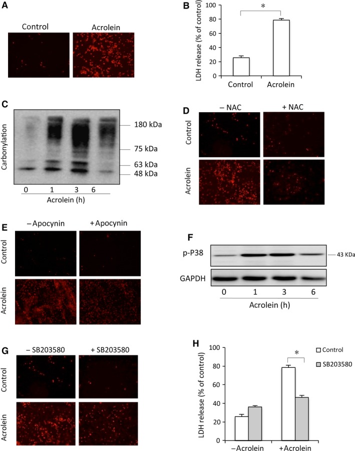
Acrolein elicits oxidative urothelial injury. (A–B) Primarily cultured urothelial cells were exposed to 100 μM acrolein for 12 hrs. Afterwards, the cells were subjected to PI staining (A) and cell supernatants were collected for LDH assay (B). Data are expressed as mean ± S.E. (n = 3). *P < 0.01. (C) Induction of protein carbonylation by acrolein. Primarily cultured urothelial cells were exposed to 100 μM acrolein for the indicated time intervals. Then, cell lysates were subjected to OxyBlot™ Protein Oxidation Detection Kit for carbonylated proteins. (D, E) Effects of antioxidant and NADPH oxidase inhibitor on cell viability. Urothelial cells were exposed to 100 μM acrolein for 12 hrs in the presence or absence of 10 mM NAC (D), or 10 mM apocynin (E). After that, cells were subjected to PI staining. (F) Induction of P38 phosphorylation by acrolein. Urothelial cells were incubated with 100 μM acrolein for the indicated time. Cellular lysates were subjected to Western blot analysis for phosphorylated P38. (G, H) Effects of P38 inhibitor SB230850 on cell viability. Urothelial cells were incubated with 100 μM acrolein for 12 hrs in the presence or absence of 20 μM P38 inhibitor SB203580. Afterwards, cells were stained with PI (G) and cell supernatants were collected for LDH assay (H). Data are expressed as mean ± S.E. (n = 3). *P < 0.01.
