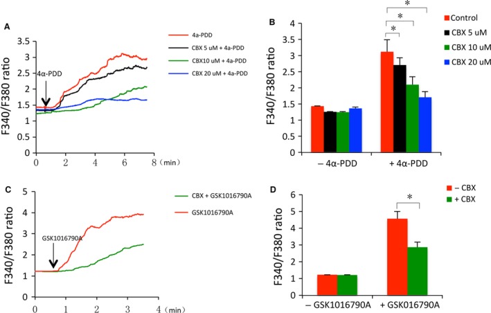
Figure 7.

CBX suppresses TRPV4 agonists‐induced increase in intracellular Ca2+. (A) Effect of CBX on 4α‐PDD‐elicited Ca2+ influx. NRK‐52E cells were pre‐treated with or without CBX for 30 min. and exposed to 3 μM 4α‐PDD in the presence or absence of CBX for the indicated time. The average levels of intracellular Ca2+ among 80–100 cells in a single study were assessed through ratiometric imaging with fura‐2 at 340 nm and 380 nm (F340/F380). The results are presented as dynamic traces of Ca2+ over time. (B) The intracellular Ca2+ level at basal and peak in (A) was quantitated. (C) Effect of CBX on GSK1016790A‐elicited Ca2+ influx. NRK‐52E cells were either pre‐treated with 10 μM CBX or left untreated for 30 min. and exposed to 5 nM GSK1016790A in the presence or absence of CBX for the indicated time. The intracellular Ca2+ measurement was performed the same as above. (D) The basal and peak intracellular Ca2+ level in (C). Data are expressed as mean ± S.E. (n = 80 ~ 100). *P < 0.01.
