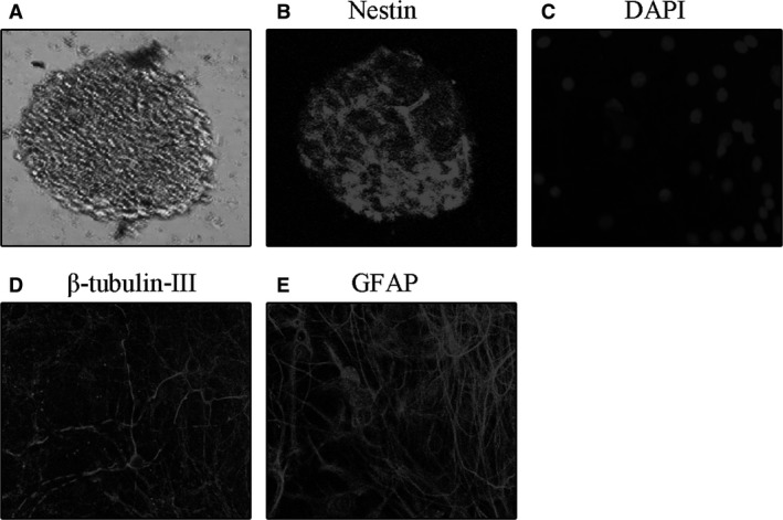
Figure 1.

The images of Proliferation and differentiation of NSCs. (A) Representative photomicrograph of neurospheres in NSC culture. (B) Immunofluorescence staining for nestin of purified NSCs. (C) Nucleus staining with DAPI of differentiated cells derived from NSCs. (D) Immunofluorescence staining for β‐tubulin‐III‐positive neurons derived from NSCs. (E) Immunofluorescence staining for GFAP‐positive protoplasmic astrocytes derived from NSCs.
