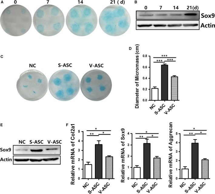
Figure 3.

Chondrogenesis of stem cells from SC adipose tissue and VS adipose tissue in micromass cultures. (A and B) S‐ASCs in micromass cultures were treated with chondrogenic media at different times. Cultures were stained with Alcian blue (A) and Western blot analysis of the lysates (40 μg) (B) (C–F) the chondrogenesis capabilities of S‐ASCs and V‐ASCs were evaluated by Alcian blue staining (C); diameter of the micromass; (D) (n = 4), Western blot analysis of lysates (40 μg); (E) and relative mRNA expression of the indicated chondrocyte‐related markers (F) (n = 3) with non‐differentiated or differentiated medium on day 21. *P < 0.05, **P < 0.01, ***P < 0.001.
