Figure 1.

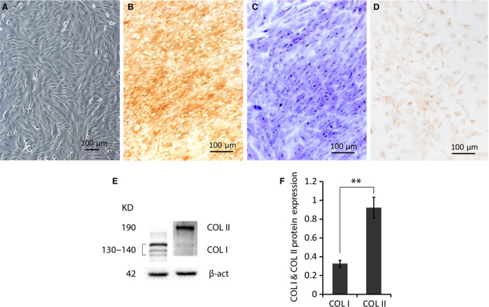
Characterization of cultured rabbit chondrocytes. (A) A typical colony morphology of the passage 3 chondrocytes showed by inverted phase contrast microscopy. (B) Chondrocytic protein COL II and (D) COL I in cultured cells were exhibited by immunohistochemical staining using specific antibodies, respectively. (C) Cellular proteoglycans were specifically stained by toluidine blue. (E) Representative Western blotting of COL I and COL II in total proteins of cultured chondrocytes using specific antibodies. (F) The plotting (n = 4) of normalized densities against the internal control β‐actin shows much higher COL II (≈3 times) than COL I (n = 4). **P < 0.01.
