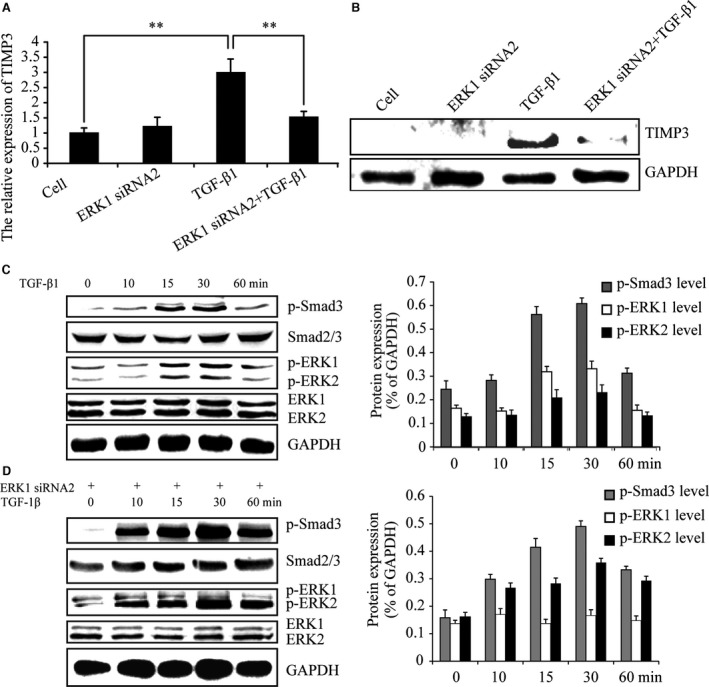
Figure 2.

ERK1 knock‐down inhibits TGF‐β1‐induced TIMP‐3. (A and B) Real‐time PCR and Western blotting indicated TGF‐β1‐induced TIMP‐3 expression was significantly suppressed by ERK1 knock‐down. (C)At the indicated time‐points, Western blotting indicated ERK1/2 and Smad3 were phosphorylated after treatment with TGF‐β1. (D) At the indicated time‐points, Western blotting revealed that p‐ERK1 was inhibited in ERK1 siRNA2 expressing cells following TGF‐β1 stimulation. The intensity levels of p‐Smad3, p‐ERK1 and p‐ERK2 were quantified. Data shown are from one representative experiment out of the three performed. ** P‐value of <0.01.
