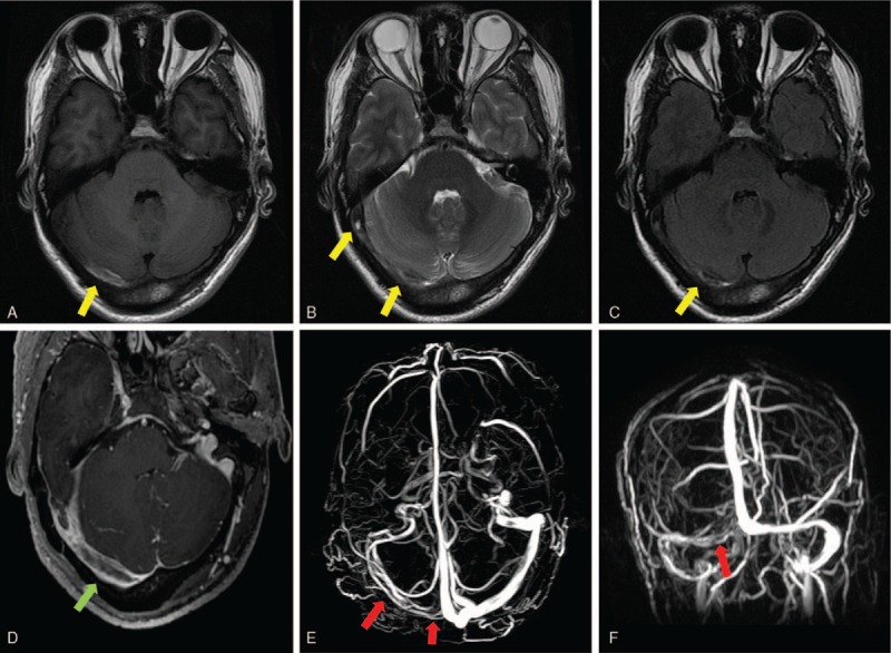
Figure 3.

Magnetic resonance imaging performed on the third day after admission showed an absence of flowing avoid in the area of right transverse sinus and sigmoid sinus (A–C). Axial sections of T1-weighted image (Fig. 4A), T2-weighted images (Fig. 4B), and fluid attenuated inversion recovery (FLAIR) images (Fig. 4C) demonstrated the presence of a hyperintense lesion on all of the 3 sequences inside the right transverse sinus, suggesting thrombosis in the right transverse sinus (A–C, yellow arrow). Axial gadolinium-enhanced T1-weighted images demonstrated filling defects in the right transverse sinus and sigmoid sinus (D, green arrow). Magnetic resonance venography demonstrated right transverse and sigmoid sinus thrombosis (E and F, red arrow).
