Figure 4.

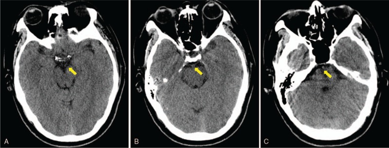
Brain computed tomographic imaging performed on day 11 provided an apparently normal image of perimesencephalic and prepontine cisterns, demonstrating a complete absorption of hemorrhage (A, B and C, yellow arrow).

Brain computed tomographic imaging performed on day 11 provided an apparently normal image of perimesencephalic and prepontine cisterns, demonstrating a complete absorption of hemorrhage (A, B and C, yellow arrow).