
Figure 4.
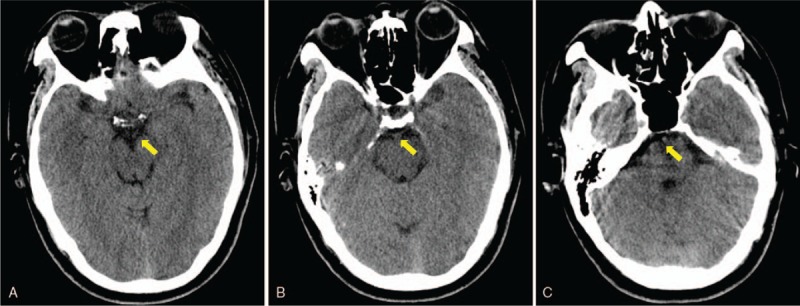
Brain computed tomographic imaging performed on day 11 provided an apparently normal image of perimesencephalic and prepontine cisterns, demonstrating a complete absorption of hemorrhage (A, B and C, yellow arrow).
Official websites use .gov
A
.gov website belongs to an official
government organization in the United States.
Secure .gov websites use HTTPS
A lock (
) or https:// means you've safely
connected to the .gov website. Share sensitive
information only on official, secure websites.
Brain computed tomographic imaging performed on day 11 provided an apparently normal image of perimesencephalic and prepontine cisterns, demonstrating a complete absorption of hemorrhage (A, B and C, yellow arrow).