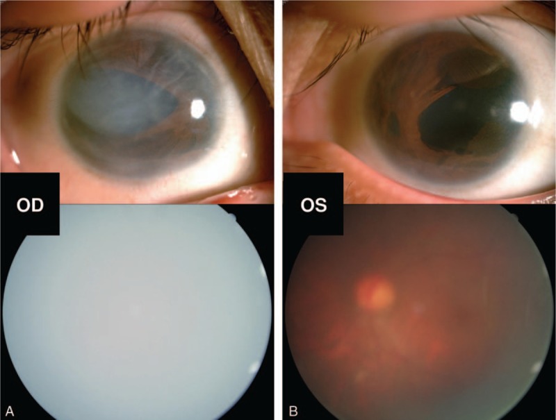
Figure 1.

Ocular traits of the recruited patient. Biomicroscopic photograph of the anterior segment showed that corneal edema with characteristic corneal posterior embryotoxon and iris changes of stromal hypoplasia with irregular-shaped pupils of her right eye (A upper). The traits of the left anterior segment included stromal hypoplasia and irregular-shaped pupil (B upper). The fundus photography of bilateral eyes was not clear because of corneal edema and cataract (A, B lower).
