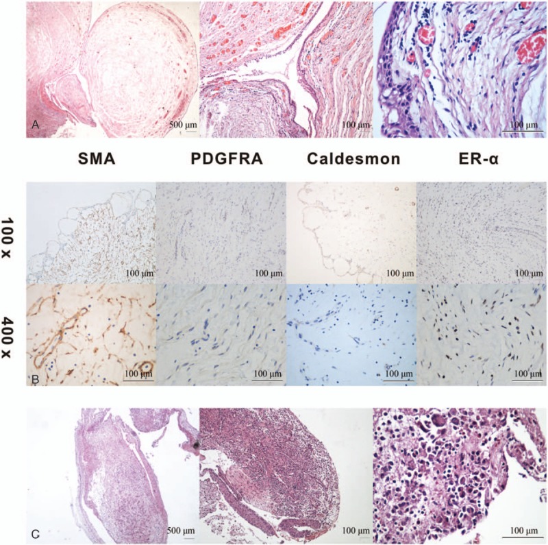
Figure 2.

(A) Hematoxylin-eosin staining of the mass covered with squamous epithelium and composed of spindle cells in a highly vascular background with an edematous and mucinous matrix with capillary vessels that were infiltrated by inflammatory cells at the original magnification ×20, ×100, and ×400. (B) Immunochemistry staining revealed moderate to strong expressions of SMA, PDGFRA, caldesmon, and ER-α in the spindle cells at magnifications of ×100 and ×400. (C) At one-month follow-up, the mucosal protrusion at the original area for hematoxylin-eosin staining of the inflammatory granulation with eosinophil and mononuclear cell infiltrations at the original magnification ×20, ×100, and ×400. Histological stains of the tissue revealed inflammatory granulation with eosinophil and mononuclear cell infiltrations. SMA = smooth muscle actin, PDGFRA = platelet-derived growth factor receptor α, ER-α = estrogen receptor-α.
