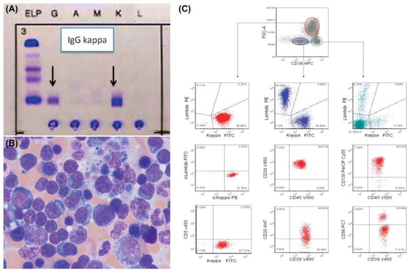
Figure 1.

(A) Serum immunofixation. (B) Bone marrow aspirate smear showing numerous lymphoplasmacytic cells. (C) Bone marrow aspirate flow cytometry analysis showing a CD38 + population (red), comprising larger than normal lymphocytes, that expresses CD19, CD138, CD45, dim CD20, CD27 and partial CD56, but is negative for CD5 and CD28. This population comprises 30% of the B-cells and is kappa immunoglobulin light chain-restricted (both surface and intracytoplasmic), most likely representing the plasmacytoid lymphocytes observed in the smear. Additionally, polyclonal B-cells (blue) and precursor B-cells (green) are present. FITC, fluorescein isothiocyanate; PE, phycoerythrin; PerCP, peridinin-chlorophyll-protein complex; APC, allophycocyanin; ic, intracytoplasmic.
