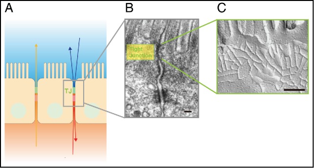
Figure 1.

Tight Junctions (TJs) are composed of claudins. (A) Schematic drawing showing the location of TJs between epithelial cells. (B) Electron micrograph of intestinal epithelial cells. TJs are located at the uppermost region of the cell-cell adhering junctional complex. Scale bar, 200 nm. (C) Freeze fracture electron micrograph of TJs. Scale bar, 200 nm. Images of (A) and (B) are modified from Tsukita et al., 20011. © Nature Reviews Molecular Cell Biology. Reproduced by permission of Nature Publishing Group. Permission to reuse must be obtained from the rightsholder.
