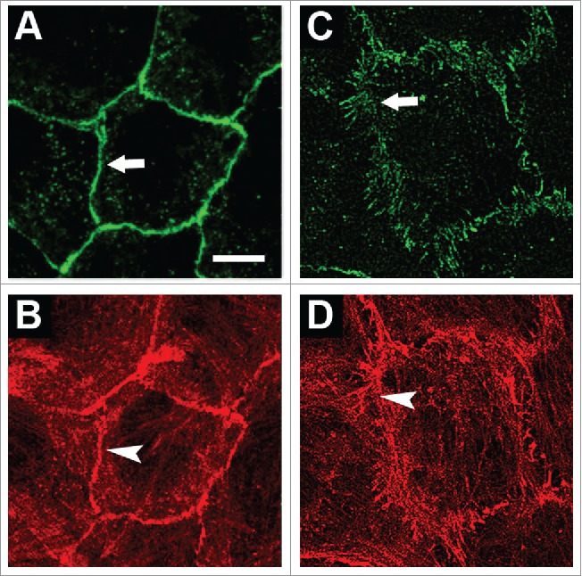
Figure 1.

Organization of AJs and the actin cytoskeleton in normal and transformed IAR cells. Cells were stained for E-cadherin (A, C) and actin (B, D). (A-B) In normal IAR-2 epithelial cells, AJs organized as adhesion belts encircling each cell and co-localizing with circumferential actin bundles. (C-D) In transformed IAR-6–1 epithelial cells, AJs organized into clusters (radial AJs) associated with thin actin bundles. © PLoS One. Reproduced by permission of Natalya Gloushankova. Permission to reuse must be obtained from the rightsholder.57
