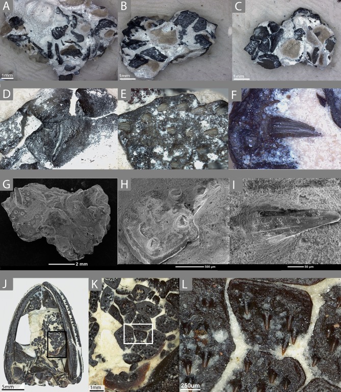
Figure 1. The external morphology of the palatal plates in the sampled specimen (ROM 76838) and Pasawioops.
(A–C) images of the sampled block of palatal plates (ROM 76838) from various views; (D) an image of the dorsal surface of the palatal plates; (E) an enlarged view of the denticulate surface of the plate; (F) an individual tooth on the plate, showing fluting; (G) an SEM image of the block from which the plates were isolated; (H) enlarged SEM image of the denticulate surface of the plate; (I) an SEM image of a single tooth; (J) an image of the palatal view of the holotype of Pasawioops (OMNH 73019); (K) An enlarged view of the palatal plates; (L) an enlarged view of the dentition on the palatal plates showing the orientation of the dentition.

