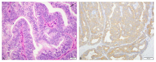
Figure 2.
Microscopic pictures of a poorly differentiated thyroid carcinoma composed of a papillary thyroid carcinoma, columnar cell phenotype with high mitotic rate (6 mitosis/10 high power fields, 400x) presenting as a 4.5 cm left neck mass in a 76 year old female. No carcinoma was found in the thyroidectomy A: Papillae lined by columnar cells with pseudostratified nuclei associated with mitosis (arrow). B: BRAF V600E immunostain was positive in the tumor.

